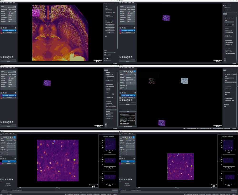
A package for 3D cell segmentation with deep learning, including a napari plugin: training, inference, and data review. In particular, this project was developed for analysis of confocal and mesoSPIM-acquired (cleared tissue + lightsheet) tissue datasets, but is not limited to this type of data. Check out our preprint for more information!
💻 See the Installation page in the documentation for detailed instructions.
📚 Documentation is available at https://AdaptiveMotorControlLab.github.io/CellSeg3D
📚 For additional examples and how to reproduce our paper figures, see: https://github.com/C-Achard/cellseg3d-figures
pip install napari_cellseg3d
To use the plugin, please run:
napari
Then go into Plugins > napari_cellseg3d, and choose which tool to use.
- Review (label): This module allows you to review your labels, from predictions or manual labeling, and correct them if needed. It then saves the status of each file in a csv, for easier monitoring.
- Inference: This module allows you to use pre-trained segmentation algorithms on volumes to automatically label cells and compute statistics.
- Train: This module allows you to train segmentation algorithms from labeled volumes.
- Utilities: This module allows you to perform several actions like cropping your volumes and labels dynamically, by selecting a fixed size volume and moving it around the image; fragment images into smaller cubes for training; or converting labels from instance to segmentation and the opposite.
The strength of our approach is we can match supervised model performance with purely self-supervised learning, meaning users don't need to spend (hundreds) of hours on annotation. Here is a quick look of our key results. TL;DR see panel f, which shows that with minmal input data we can outperform supervised models:
Figure 1. Performance of 3D Semantic and Instance Segmentation Models. a: Raw mesoSPIM whole-brain sample, volumes and corresponding ground truth labels from somatosensory (S1) and visual (V1) cortical regions. b: Evaluation of instance segmentation performance for baseline thresholding-only, supervised models: Cellpose, StartDist, SwinUNetR, SegResNet, and our self-supervised model WNet3D over three data subsets. F1-score is computed from the Intersection over Union (IoU) with ground truth labels, then averaged. Error bars represent 50% Confidence Intervals (CIs). c: View of 3D instance labels from supervised models, as noted, for visual cortex volume in b evaluation. d: Illustration of our WNet3D architecture showcasing the dual 3D U-Net structure with our modifications.
Read the article here !
-
v0.2.2:
- Updated the Colab Notebooks for training and inference
- New models available in the inference demo notebook
- CRF optional post-processing adjustments (and pip install directly)
-
v0.2.1:
- Updated plugin default behaviors across the board to be more readily applicable to demo data
- Threshold value in inference is now automatically set by default according to performance on demo data on a per-model basis
- Added a grid search utility to find best thresholds for supervised models
-
v0.2.0:
- Changed project name to "napari_cellseg3d" to avoid setuptools deprecation
- Small API changes for training/inference from a script
- Some fixes to WandB integration and csv saving after training
Previous additions:
- v0.1.2: Fixed manifest issue for PyPi
- Improved training interface
- Unsupervised model : WNet3D
- Generate labels directly from raw data!
- Can be trained in napari directly or in Google Colab
- Pretrained weights for mesoSPIM whole-brain cell segmentation
- WandB support (install wandb and login to use automatically when training)
- Remade and improved documentation
- Moved to Jupyter Book
- Dedicated installation page, and working ARM64 install for macOS Silicon users
- New utilities
- Many small improvements and many bug fixes
Compatible with Python 3.8 to 3.10. Requires napari, PyTorch and MONAI. Compatible with Windows, MacOS and Linux. Installation of the plugin itself should not take more than 30 minutes, depending on your internet connection, and whether you already have Python and a package manager installed.
For PyTorch, please see the PyTorch website for installation instructions.
A CUDA-capable GPU is not needed but very strongly recommended, especially for training.
If you get errors from MONAI regarding missing readers, please see MONAI's optional dependencies page for instructions on getting the readers required by your images.
Please reach out if you have any issues with the installation, we will be happy to help!
To avoid issues when installing on the ARM64 architecture, please follow these steps.
-
Create a new conda env using the provided conda/napari_CellSeg3D_ARM64.yml file :
git clone https://github.com/AdaptiveMotorControlLab/CellSeg3d.git cd CellSeg3d conda env create -f conda/napari_CellSeg3D_ARM64.yml conda activate napari_CellSeg3D_ARM64 -
Install a Qt backend (PySide or PyQt5)
-
Launch napari, the plugin should be available in the plugins menu.
Help us make the code better by reporting issues and adding your feature requests!
If you encounter any problems, please file an issue along with a detailed description.
You can generate docs locally by running make html in the docs/ folder.
Before testing, install all requirements using pip install napari-cellseg3d[test].
pydensecrf is also required for testing.
To run tests locally:
- Locally : run
pytest napari_cellseg3d\_testsin the plugin folder. - Locally with coverage : In the plugin folder, run
coverage run --source=napari_cellseg3d -m pytestthencoverage xmlto generate a .xml coverage file. - With tox : run
toxin the plugin folder (will simulate tests with several python and OS configs, requires substantial storage space)
Contributions are very welcome.
Please ensure the coverage at least stays the same before you submit a pull request.
For local installation from Github cloning, please run:
pip install -e .
Distributed under the terms of the MIT license.
"napari-cellseg3d" is free and open source software.
@article {10.7554/eLife.99848,
article_type = {journal},
title = {CellSeg3D, Self-supervised 3D cell segmentation for fluorescence microscopy},
author = {Achard, Cyril and Kousi, Timokleia and Frey, Markus and Vidal, Maxime and Paychere, Yves and Hofmann, Colin and Iqbal, Asim and Hausmann, Sebastien B and Pagès, Stéphane and Mathis, Mackenzie Weygandt},
editor = {Cardona, Albert},
volume = 13,
year = 2025,
month = {jun},
pub_date = {2025-06-24},
pages = {RP99848},
citation = {eLife 2025;13:RP99848},
doi = {10.7554/eLife.99848},
url = {https://doi.org/10.7554/eLife.99848},
abstract = {Understanding the complex three-dimensional structure of cells is crucial across many disciplines in biology and especially in neuroscience. Here, we introduce a set of models including a 3D transformer (SwinUNetR) and a novel 3D self-supervised learning method (WNet3D) designed to address the inherent complexity of generating 3D ground truth data and quantifying nuclei in 3D volumes. We developed a Python package called CellSeg3D that provides access to these models in Jupyter Notebooks and in a napari GUI plugin. Recognizing the scarcity of high-quality 3D ground truth data, we created a fully human-annotated mesoSPIM dataset to advance evaluation and benchmarking in the field. To assess model performance, we benchmarked our approach across four diverse datasets: the newly developed mesoSPIM dataset, a 3D platynereis-ISH-Nuclei confocal dataset, a separate 3D Platynereis-Nuclei light-sheet dataset, and a challenging and densely packed Mouse-Skull-Nuclei confocal dataset. We demonstrate that our self-supervised model, WNet3D – trained without any ground truth labels – achieves performance on par with state-of-the-art supervised methods, paving the way for broader applications in label-scarce biological contexts.},
keywords = {self-supervised learning, artificial intelligence, neuroscience, mesoSPIM, confocal microscopy, platynereis},
journal = {eLife},
issn = {2050-084X},
publisher = {eLife Sciences Publications, Ltd},
}
This plugin was developed by originally Cyril Achard, Maxime Vidal, Mackenzie Mathis. This work was funded, in part, from the Wyss Center to the Mathis Laboratory of Adaptive Intelligence. Please refer to the documentation for full acknowledgements.
This napari plugin was generated with Cookiecutter using @napari's cookiecutter-napari-plugin template.


